Protecting healthcare
workers from needlestick injuries
Non-Healing Rib Fractures: Signs Your Injury Needs More Than Rest
Rib fractures are among the most common injuries seen after blunt chest trauma, whether from a car accident, a hard fall, a sports collision, or even a forceful bout of coughing in people with weakened bones. Most of the time, a cracked or broken rib heals on its own within six to twelve weeks. The standard advice, rest, careful movement, and adequate pain relief, works well for straightforward cases. The bones knit back together, the pain fades, and life resumes. For a meaningful portion of people, though, that process stalls or fails entirely, leaving them with an injury that never quite resolves.
When a rib fracture stops progressing toward healing, the consequences reach well beyond ongoing discomfort. Persistent chest wall instability can limit breathing, disrupt sleep, and prevent a return to normal activity for months or even years. The problem is that many people assume lingering pain is simply part of the process, or that their body just needs more time. Understanding what a truly non-healing rib fracture looks and feels like, and what drives that outcome, is the first step toward getting the right care.
How Rib Fractures Normally Heal and Why the Process Sometimes Breaks Down
Bone healing is a biological sequence that moves through several overlapping phases. In the first days after a fracture, a clot forms at the break site, and the body begins recruiting cells that will eventually lay down new bone tissue. Over the following weeks, a soft callus of fibrous material and cartilage bridges the gap. That callus gradually mineralizes and hardens into true bone, a process that typically reaches structural completion within six to twelve weeks for most rib fractures.
The rib cage presents particular challenges for this process. Each rib moves with every breath, meaning the fractured ends experience repeated micro-motion that can disrupt the developing callus. A single, non-displaced fracture in an otherwise healthy person usually has enough stability to heal despite this movement. Displaced fractures, where the broken ends are shifted out of alignment, and fractures involving multiple adjacent ribs create far more instability, which raises the risk of prolonged or failed healing.
Medically, a fracture is classified as having delayed union when it has not achieved bony continuity by six months after the injury. If healing fails altogether and the bone ends form a fibrous false joint instead of fusing, the condition is called a nonunion, and it is permanent unless surgically corrected. A third scenario, malunion, occurs when the bone heals but in a misaligned position, distorting the chest wall and creating chronic mechanical pain.
Nonunion affects approximately 12 percent of rib fractures in patients who sustain three or more fractured ribs, according to research published in the journal Injury. That figure rises significantly when fractures are displaced or when other risk factors are present. The condition is more common than many patients and even some clinicians realize, partly because standard follow-up after rib injuries is often limited to a single chest X-ray that may not capture early signs of healing failure.
Red Flags That Signal Your Fracture Is Not Progressing
The clearest warning sign is pain that does not follow the expected trajectory. A healing rib fracture should feel progressively less painful over weeks. Pain that remains intense beyond six to eight weeks, that returns after a period of improvement, or that is sharply localized at the fracture site rather than diffuse suggests the bone is not consolidating properly.
A clicking or popping sensation in the chest is particularly telling. This occurs when the broken bone ends are mobile enough to shift against each other during movement or breathing, a phenomenon patients often describe as a grinding or jabbing sensation. It typically means the fracture site lacks the stability needed for healing and has likely developed fibrous nonunion tissue. Unlike the general soreness of a healing rib, this mechanical symptom does not respond to rest and often worsens with activity.
Breathlessness that persists or worsens over time also deserves prompt attention. When a rib fracture fails to heal, the affected portion of the chest wall may remain abnormally mobile, impairing the chest's ability to expand effectively during inhalation. Shallow breathing caused by pain allows mucus to accumulate in the lower airways, raising the risk of repeated chest infections. A tender lump or visible deformity at the fracture site that does not diminish over several weeks is a further sign that something beyond normal healing is occurring.
Risk Factors That Make Non-Healing More Likely
Smoking is one of the most consistently documented contributors to fracture nonunion. Nicotine impairs osteoblasts, the cells responsible for building new bone, while reducing blood flow to healing tissue. Research has found that smokers are more than three times more likely to experience complications in bone healing than non-smokers, and this association applies directly to rib fractures.
Osteoporosis elevates the risk significantly. In osteoporotic bone, the structural framework that supports callus formation is already compromised, and the initial fracture may have occurred from a relatively low-energy impact, suggesting poor baseline bone quality. Older adults, particularly post-menopausal women, are disproportionately represented among patients who develop rib nonunion. Nutritional deficiencies, especially low calcium, vitamin D, and protein intake, deprive the body of essential materials for bone repair. Diabetes impairs both circulation and immune function, and prolonged corticosteroid use has a well-established inhibitory effect on bone metabolism.
Displacement of the fracture fragments matters greatly. When broken ends overlap or are significantly separated, the gap they create is too large for callus to bridge reliably without mechanical support. Fractures in which the same rib is broken at more than one point, known as comminuted fractures, are particularly prone to instability and nonunion. There is also growing evidence from meta-analyses that regular use of nonsteroidal anti-inflammatory drugs (NSAIDs) after a fracture may impair healing by suppressing the inflammatory signals that initiate bone repair, a complicating factor since NSAIDs are among the most commonly prescribed medications for rib pain.
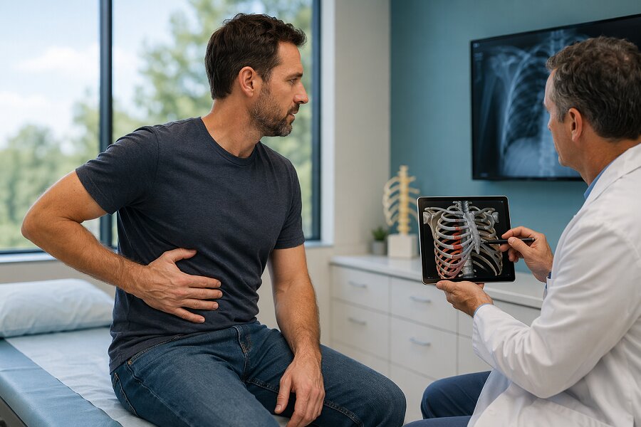
Diagnosis, Imaging, and When Surgery Becomes the Right Path
A standard chest X-ray, useful for identifying fractures at the time of injury, often undersells the severity of the problem. Hairline fractures may be entirely invisible on plain films, and early signs of nonunion, such as absent callus formation or a persistent fracture line at three months, can be subtle. A CT scan provides far greater detail, clearly showing fracture displacement, callus development, and any associated complications such as fluid in the chest cavity.
When CT imaging confirms nonunion or significant instability several months after the original injury, surgical stabilization becomes a serious consideration, particularly when conservative measures have failed. The most established approach is open reduction and internal fixation (ORIF), in which the surgeon realigns the fractured rib segments and secures them with anatomically contoured titanium plates and screws. These implants match the natural curve of the ribs and allow the bone to heal correctly while the chest continues to move with breathing.
Outcomes following surgery for rib nonunion are generally favorable. Published case series show meaningful reductions in pain and improved respiratory function in the majority of patients who undergo fixation. Most patients begin experiencing relief from mechanical symptoms, particularly the clicking and instability, within weeks of the procedure. The decision to operate always involves weighing the duration and severity of symptoms against individual health factors and the patient's goals for recovery.
When Waiting Is No Longer the Right Answer
The gap between what patients experience and what gets acted upon is a real problem with rib injuries. Because initial treatment is nearly always conservative, and many patients are advised simply to wait, those who do not heal as expected can spend months without a clear explanation or a path forward. If pain is still significantly affecting your breathing, sleep, or daily activity beyond two to three months, or if you are experiencing clicking, instability, or persistent breathlessness, a reassessment by a chest wall specialist or thoracic surgeon is warranted. A repeat CT scan at that stage can clarify exactly what is happening at the fracture site and guide a more informed conversation about whether continued rest is reasonable or whether intervention will produce better results meaningfully.